Understanding Animal Cell Structure
Animal cell coloring sheet answers – The animal cell, a fundamental building block of animal life, is a complex and fascinating entity. Its intricate internal organization allows for the efficient performance of numerous life-sustaining processes. Understanding its structure is key to comprehending the mechanisms of life itself. This exploration will delve into the major components of the animal cell, their individual functions, and how they differ from their plant cell counterparts.
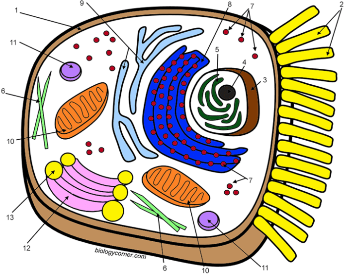
Major Organelles and Their Functions, Animal cell coloring sheet answers
The animal cell houses a variety of specialized organelles, each performing a specific role in maintaining cellular homeostasis. These organelles work in concert, creating a dynamic and interdependent system.
| Organelle | Function | Description | Example |
|---|---|---|---|
| Nucleus | Houses genetic material (DNA); controls cell activities. | A large, membrane-bound organelle containing chromosomes. | The control center, dictating protein synthesis and cell division. |
| Ribosomes | Synthesize proteins. | Small, granular structures, either free-floating or attached to the endoplasmic reticulum. | Essential for building enzymes and structural proteins. |
| Endoplasmic Reticulum (ER) | Synthesizes lipids and proteins; transports materials within the cell. | A network of interconnected membranes; Rough ER (with ribosomes) and Smooth ER (without ribosomes). | The cell’s internal highway system. |
| Golgi Apparatus | Processes and packages proteins for secretion or use within the cell. | A stack of flattened sacs (cisternae). | The cell’s post office, modifying and sorting proteins. |
| Mitochondria | Generate energy (ATP) through cellular respiration. | Double-membrane bound organelles, often called the “powerhouses” of the cell. | Essential for providing energy for cell functions. |
| Lysosomes | Break down waste materials and cellular debris. | Membrane-bound sacs containing digestive enzymes. | The cell’s recycling and waste disposal system. |
| Cell Membrane | Regulates the passage of substances into and out of the cell. | A selectively permeable membrane surrounding the cell. | Maintains the cell’s internal environment. |
Differences Between Plant and Animal Cells
Plant and animal cells share many similarities, but key distinctions exist. These differences reflect the unique needs and functions of each cell type.Plant cells possess a rigid cell wall providing structural support and protection, absent in animal cells. Plant cells also contain chloroplasts, the sites of photosynthesis, enabling them to produce their own food, a capability lacking in animal cells.
Need help with your animal cell coloring sheet answers? Understanding cell structures can be challenging, but a fun way to learn is by visualizing. For a broader approach to animal anatomy, check out this fantastic resource for animals coloring book pdf free download , which can complement your cell studies. Returning to those animal cell coloring sheet answers, remember to focus on the key organelles like the nucleus and mitochondria for accurate completion.
Finally, plant cells typically have a large central vacuole for water storage and maintaining turgor pressure, while animal cells have smaller, less prominent vacuoles.
Coloring Sheet Content Analysis

Analyzing the content of animal cell coloring sheets offers a valuable opportunity to assess the understanding and visual representation of cellular structures. These sheets, often used in educational settings, provide a simplified yet potentially informative depiction of complex biological systems. The accuracy and completeness of these representations vary considerably.
A typical animal cell coloring sheet will usually depict a selection of key organelles. The specific organelles included may vary depending on the educational level and the focus of the sheet, but common inclusions are the nucleus, cytoplasm, cell membrane, mitochondria, ribosomes, endoplasmic reticulum (both rough and smooth), Golgi apparatus, lysosomes, and sometimes vacuoles (though these are generally smaller and less prominent in animal cells compared to plant cells).
Organelle Depictions on Coloring Sheets
Coloring sheets often employ simplified visual representations to facilitate understanding. For example, the nucleus is typically shown as a large, centrally located, circular or oval structure. Mitochondria are frequently depicted as bean-shaped or sausage-shaped structures scattered throughout the cytoplasm. The endoplasmic reticulum is often illustrated as a network of interconnected tubules and sacs, while the Golgi apparatus is frequently represented as a stack of flattened sacs or cisternae.
Ribosomes, due to their small size, are often shown as tiny dots associated with the endoplasmic reticulum or scattered in the cytoplasm. The cell membrane is usually depicted as a single line outlining the cell’s boundary. Lysosomes may be shown as small, circular structures containing darker material, symbolizing their role in waste breakdown.
Comparison of Organelle Representations Across Different Coloring Sheets
Variations in the visual representation of organelles can be observed across different coloring sheets. Some may use more detailed illustrations, showing internal structures within organelles, while others maintain a simpler, schematic approach. Color coding may also differ, with some sheets using consistent colors for specific organelles across the entire sheet, while others employ a more varied palette. The relative sizes of organelles might also vary, with some sheets accurately reflecting the proportional sizes (as much as possible in a simplified drawing), while others might exaggerate the size of certain organelles for clarity.
For instance, the nucleus is frequently shown larger than it would be proportionally, for emphasis on its importance.
Accuracy of Organelle Size and Position
The accuracy of organelle size and position on coloring sheets is often compromised for the sake of clarity and simplicity. While the general location of organelles (e.g., nucleus centrally located) is usually correct, the precise relative sizes and positions may not perfectly mirror reality. For example, the extensive network of the endoplasmic reticulum and the intricate structure of the Golgi apparatus are significantly simplified.
Furthermore, the dynamic nature of organelles, such as the constant movement and fusion of vesicles, is not typically captured in static coloring sheet illustrations. A realistic representation would require a far more complex illustration, making it less suitable for basic educational purposes. Consider a coloring sheet showing the nucleus taking up half the cell; this is inaccurate, but serves a didactic purpose of highlighting the organelle’s importance.
Educational Value of Coloring Sheets: Animal Cell Coloring Sheet Answers

Coloring sheets, often underestimated, offer a surprisingly powerful pedagogical tool, particularly in the realm of science education. Their engaging nature transcends the simple act of coloring, fostering active learning and deeper comprehension of complex concepts. The visual and kinesthetic elements involved contribute significantly to knowledge retention and understanding. In the context of biology, specifically animal cell structure, coloring sheets prove exceptionally valuable.The act of coloring an animal cell diagram transforms passive learning into an active process.
Students aren’t simply reading about the cell’s components; they are actively engaging with them, associating names with visual representations. This multi-sensory approach enhances memory encoding and retrieval. The detailed coloring process encourages careful observation of the different organelles and their relative sizes and locations within the cell. This visual reinforcement strengthens understanding and improves long-term retention of complex information.
Moreover, the hands-on nature of the activity makes learning more enjoyable and less daunting, thereby increasing student engagement and motivation.
Enhanced Memorization Through Visual Association
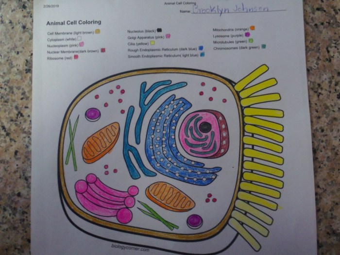
Coloring sheets facilitate memorization by creating strong visual associations between the names of cell organelles and their physical representations. For example, coloring the nucleus purple and the mitochondria blue helps students easily recall the location and function of each organelle. This visual cueing system leverages the brain’s natural capacity for visual learning, resulting in more effective memorization compared to rote learning from textbooks alone.
The act of repeatedly writing the names of the organelles while coloring further strengthens the connections between the visual and verbal representations. This combination of visual and kinesthetic activities significantly improves the memorization process. Studies have shown that incorporating visual aids into learning materials significantly improves retention rates, and coloring sheets provide an effective and engaging way to achieve this.
Lesson Plan Incorporating an Animal Cell Coloring Sheet Activity
This lesson plan uses an animal cell coloring sheet to teach students about the structure and function of animal cells.Pre-Activity Assessment: A brief quiz assessing prior knowledge of cells and their components. Example questions could include: “What is a cell?” or “What are some of the parts of a cell you already know?”Activity: Students are given a pre-printed animal cell coloring sheet with labeled organelles.
They color each organelle a different color, referencing a key that provides the name and a brief description of each organelle’s function. This coloring process is followed by a short discussion where students can share what they have learned.Post-Activity Assessment: A worksheet with diagrams of animal cells, some with organelles labeled and others without. Students label the unlabeled diagrams and answer short answer questions about the function of different organelles.
This could also include a short matching activity, pairing the name of an organelle with its function. This assessment gauges the effectiveness of the coloring sheet activity in improving understanding and retention of information. A higher percentage of correct answers on the post-activity assessment compared to the pre-activity assessment demonstrates the effectiveness of this teaching method.
Creating an Enhanced Coloring Sheet
Elevating the simple act of coloring into a powerful learning experience requires a meticulously designed coloring sheet. This enhanced version will not only engage students visually but also foster a deeper understanding of animal cell structures and their functions. We aim to create a more accurate and detailed representation, moving beyond basic shapes to a more realistic portrayal of the organelles within an animal cell.An enhanced animal cell coloring sheet utilizes color not merely for aesthetic appeal, but as a powerful tool to visually differentiate the various organelles and their functions.
Strategic color choices will highlight the unique roles each component plays in maintaining cellular life. This will enable a more intuitive grasp of the cell’s complex inner workings.
Organelle Descriptions and Functions
The following table details the key organelles found within an animal cell, their functions, and suggested colors for representation on the enhanced coloring sheet. Accurate depiction is crucial for effective learning. Remember, the colors are suggestions; feel free to adapt them based on your preferences or available resources.
| Organelle | Function | Suggested Color |
|---|---|---|
| Nucleus | Contains the cell’s genetic material (DNA) and controls cell activities. | Dark Purple |
| Nucleolus | Produces ribosomes. | Light Purple |
| Ribosomes | Synthesize proteins. | Dark Blue |
| Endoplasmic Reticulum (ER) | Network of membranes involved in protein and lipid synthesis; Rough ER (with ribosomes) and Smooth ER (without ribosomes). | Rough ER: Light Blue; Smooth ER: Light Green |
| Golgi Apparatus (Golgi Body) | Processes and packages proteins and lipids. | Yellow |
| Mitochondria | Generate energy (ATP) through cellular respiration. | Red |
| Lysosomes | Break down waste materials and cellular debris. | Orange |
| Vacuoles | Store water, nutrients, and waste products; generally smaller in animal cells than in plant cells. | Light Orange |
| Cytoskeleton | Provides structural support and facilitates cell movement. | Light Gray |
| Cell Membrane | Encloses the cell and regulates the passage of substances. | Dark Green |
| Centrioles | Involved in cell division. | Brown |
Color-Coding Cellular Functions
The strategic use of color enhances the learning experience. For example, using shades of blue for protein synthesis (ribosomes and rough ER) visually connects these organelles and their shared function. Similarly, using warm colors (red and orange) for energy production (mitochondria) and waste breakdown (lysosomes) creates a visual association between these processes. The choice of green for the cell membrane emphasizes its role as a boundary and regulator.
This systematic color-coding facilitates the understanding of functional relationships between organelles.
